Open datasets
We build AI tools and help create open datasets for use by neuroscience research groups worldwide. These freely accessible datasets, built with AI and in partnership with the broader scientific community, support ongoing experiments to better understand how brains function.
A browsable petascale reconstruction of the human cortex
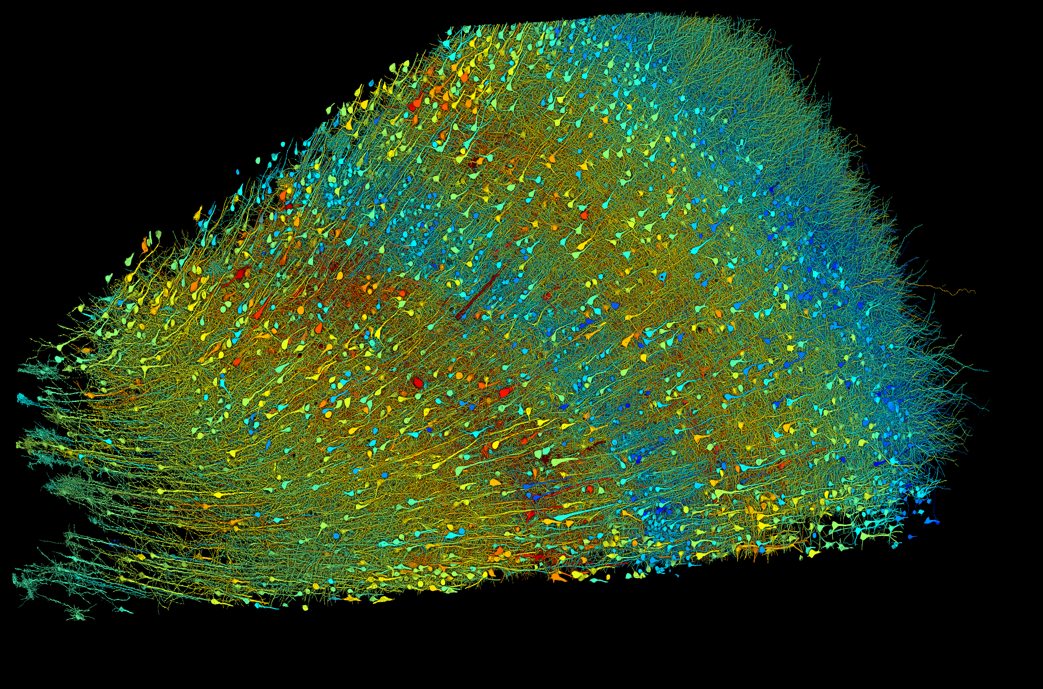
Together with the Lichtman laboratory at Harvard University, in 2021 we released the preliminary “H01” dataset, with the final version resulting in publication in Science. H01 is a 1.4 petabyte representation of a small sample of human brain tissue. Nanoscale resolution images were taken using electron microscopy, reconstructed into 3D structures and annotated using automated techniques, and then manually analyzed for insights into the structure of the human cortex. The sample covers roughly 1 cubic mm and incorporates roughly 57,000 cells, including 16,000 neurons, 32,000 glia and 8,000 blood vessel cells, as well as 150 million synapses. All are accessible and browsable within the Neuroglancer interface.
Explore the dataset.

The excitatory neurons in a 3 mm wide sample of human brain tissue, shaded according to the size of the neuron’s cell bodies, the central core.
Male fruit fly brain and central nervous system (CNS) connectome
With the Howard Hughes Medical Institute-Janelia and other partners, we present the first finalized connectome of an entire male fruit fly’s central nervous system, encompassing the central brain, optic lobes (analogous to mammalian retina) and the ventral nerve cord (analogous to the spinal cord), totaling more than 166,000 neurons. After building several partial fruit fly brain maps, this final male fruit fly connectome complements the female fruit fly connectome and provides a foundational resource for this model organism in biology.
Explore the dataset.

A small subset of cells in the male fruit fly’s brain and central nervous system.
Fish1 atlas: Whole-brain structural dataset for a larval zebrafish
This is, at the time of its release, the largest whole-brain dataset for a vertebrate. This effort led by Harvard University integrates light and electron microscopy imagery from a 7-day-old larval zebrafish. This atlas spans the brain and anterior spinal cord with over 180,000 segmented soma, 40,000 molecularly annotated neurons, and 30 million synapses. While not a comprehensive brain map of the zebrafish, this resource includes several complete circuits that represent distinct neural pathways, providing a single resource for diverse neuroscience experiments on a vertebrate.
Explore the dataset.
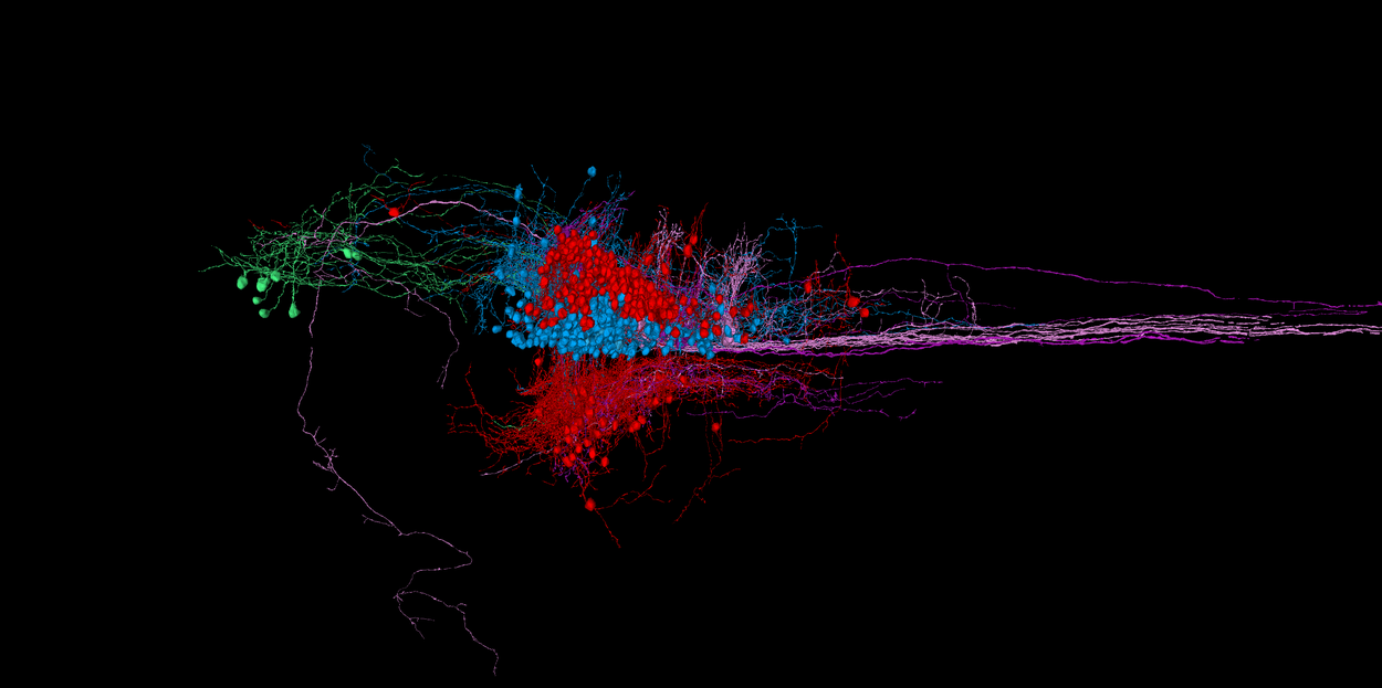
Neural circuit for the tegmental excitatory nucleus in the larval zebrafish.
Fish Fire&Wire
Our team and partners at HHMI Janelia and Harvard released a single-synapse-resolution wiring diagram for an entire larval zebrafish brain – the first complete connectome for a vertebrate. The draft version was finished in 2026. The same fish’s neuronal activity was previously recorded while demonstrating a series of behaviors. Superimposing these two datasets now allows a combined view of “firing and wiring” in a vertebrate brain. Moreover, because the dataset includes sensory structure – like nerve cells in the ears, nose and skin – as well as muscle outputs, it supports mechanistic brain models that connect environmental stimuli all the way to resulting actions.
Explore the dataset.
Individual neurons mapped in a larval zebrafish. This same fish’s neuronal activity was previously recorded while exhibiting a set of typical behaviors.
ZAPBench: Zebrafish activity prediction benchmark
Can we predict patterns of brain activity in a zebrafish? This dataset provides a foundational resource to reach that goal. In collaboration with HHMI Janelia and Harvard, ZAPBench provides a cellular-level resolution, whole-brain dataset of brain activity in a zebrafish during experiments that simulate common situations. This benchmark will support the creation of more advanced predictive models for brain activity in zebrafish – and, perhaps someday, other vertebrates. The dataset includes recordings of electrical activity of over 70,000 zebrafish neurons. This same fish specimen is currently undergoing synaptic-level anatomical mapping, so in future, it will be possible to interpret electrical activity in the context of precise neural structure.
Explore the dataset.
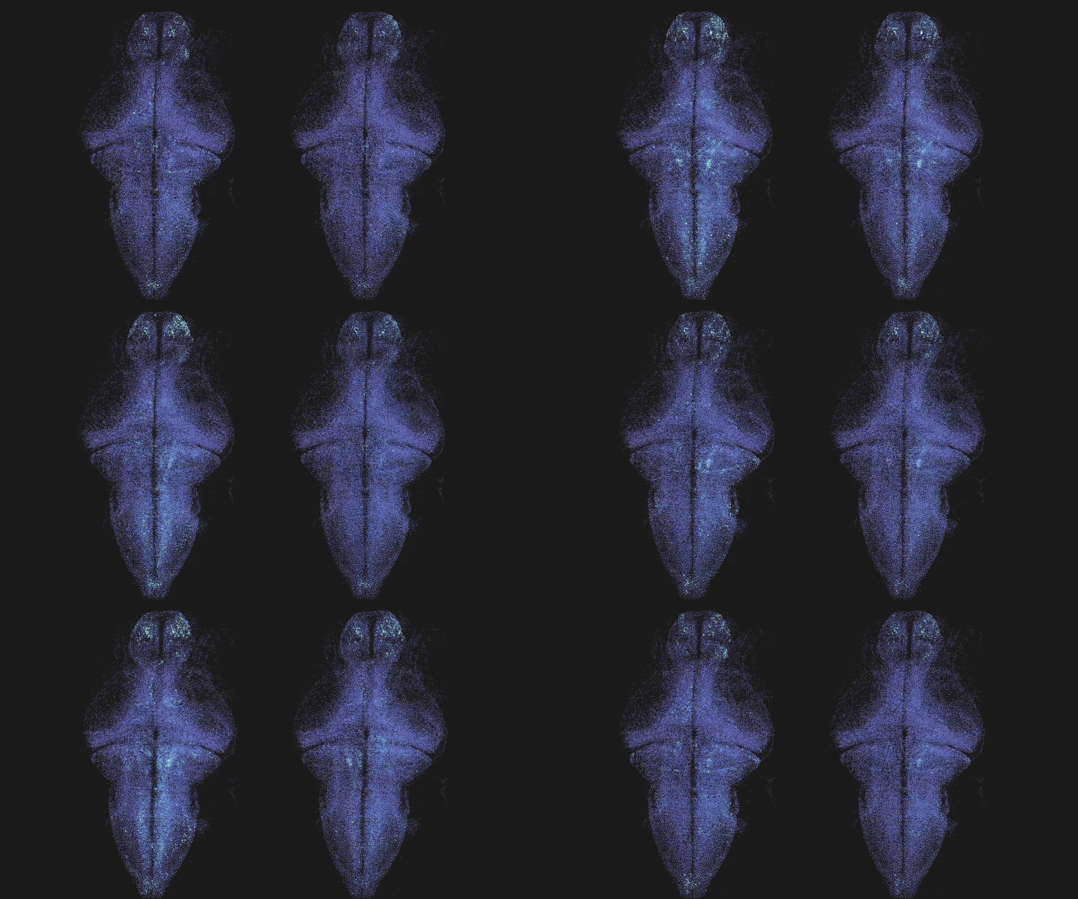
Side by side comparison of a brain activity map. Left: Ground truth. Right: Prediction.