Neural mapping
Google Research is driving progress toward precisely mapping the connections between every cell in the brain.
What is connectomics?
The human brain is perhaps the most computationally complex machine in existence, consisting of networks of billions of cells. Researchers currently don’t understand the full picture of how glitches in its network machinery contribute to mental illnesses and other diseases, such as dementia. However, the emerging connectomics field, which aims to precisely map the connections between every cell in the brain, could help solve that problem.
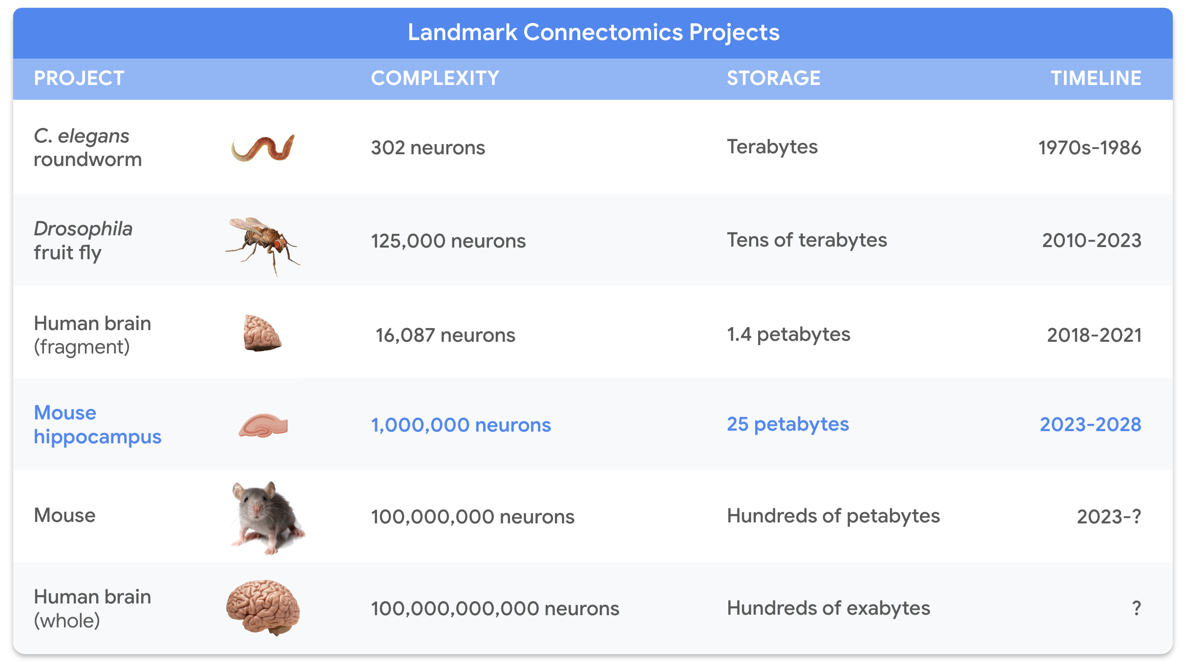
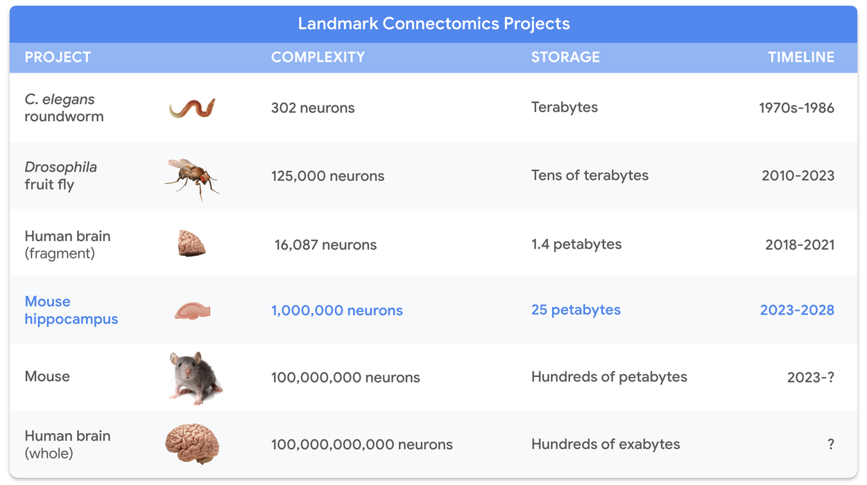
The Connectomics team at Google Research has played a key role in advancing the connectonomics field by developing new technologies that have accelerated scientific progress. These technologies enabled us to map parts of the fruit fly, mouse and human brain, and could one day help us better understand how the human brain works and how to treat brain diseases. The timeline and chart below demonstrate how connectomics has evolved since the 1970s.
Explore our recent research

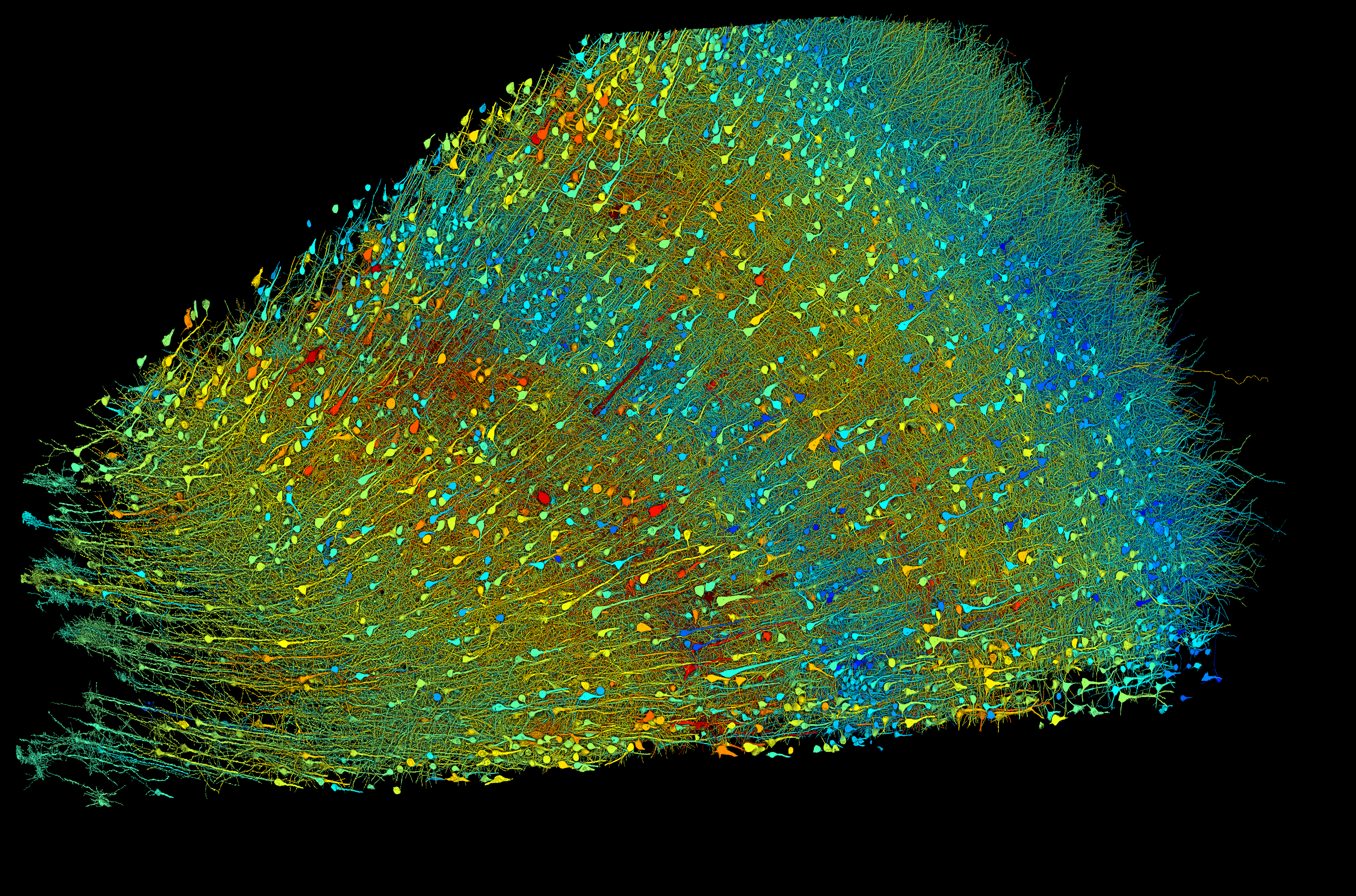
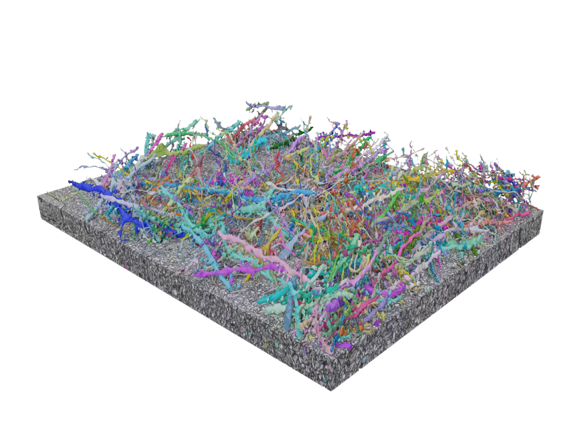
This project mapped a fragment of human brain tissue the size of half a grain of rice, incorporating roughly 16,000 neurons and 150 million synapses, that required 1.4 million gigabytes to encode. This collaborative effort revealed never-before-seen structures within the human brain.

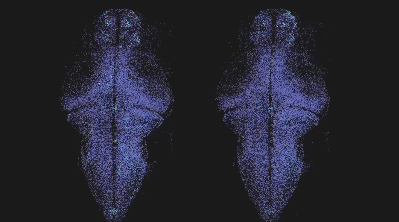
With our partners we released ZAPBench, a whole-coverage map of electrical activity in a zebrafish brain. This new benchmark will support future neuroscience experiments involving this vertebrate model organism.
Connectomics has mostly relied on costly electron microscope images. LICONN introduces a way to use widely available light microscopes to map individual neurons.
Synthetic data is commonly used to train AI models. We used AI-generated synthetic neural shapes to train an AI brain reconstruction tool, producing a measurable improvement on the state of the art.
Collaborations with the research community
A photo of Gerry Rubin. Photo credit: Matt Staley, HHMI’s Janelia Research Campus
More than a "mass of spaghetti"Gerry Rubin, director of the Howard Hughes Medical Institute's Janelia Research Campus, and a team of researchers constructed the fruit fly connectome, revolutionizing brain mapping.

A photo of Gwyneth Card. Photo credit: John Abbott, Columbia's Zuckerman Institute
Escaping dangerGwyneth Card, a neuroscientist who leads a lab at Columbia University's Zuckerman Institute, researches how insect brains are wired to help them flee from predators.
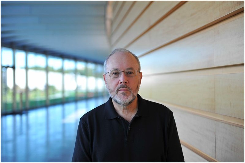
A photo of Jeff Lichtman.
The mouse connectome moonshotJeff Lichtman, a neuroscientist who leads the Lichtman Lab at Harvard University, is working towards mapping the first whole mammalian brain.
Featured publications